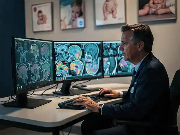
Brain MRI is the gold standard imaging test for cerebral palsy. It shows the injury directly — where in the brain, how extensive, and often when it happened. The pattern on MRI usually maps to the type of CP a child will have, making the imaging report one of the most important pieces of the diagnostic puzzle.
Of children with CP have detectable findings on MRI
No radiation
Uses magnetic fields, safe for repeat imaging in children
When pediatric neurologists confirm a cerebral palsy diagnosis, brain MRI is almost always part of the picture. About 80% of children with CP have detectable abnormalities on MRI — and the specific pattern usually does more than confirm the diagnosis. It often shows when the injury occurred, what type of CP to expect, and what other concerns might come along with the motor problems. For many families, the MRI report is the first concrete information they get about their child’s injury.
MRI is unique among imaging tools because it shows brain tissue in detail without radiation. For children — especially infants whose brains are still developing — that combination of safety and detail is exactly what CP diagnosis requires.
The reason MRI sits at the center of CP imaging isn’t mystique — it’s physics. Magnetic fields and radio waves can produce images that distinguish white matter from gray matter, identify millimeter-sized lesions, and reveal subtle structural malformations. CT and ultrasound, while useful in specific situations, simply can’t match that resolution. And because MRI uses no ionizing radiation, it can be repeated as needed without cumulative exposure concerns.
Why MRI is crucial for cerebral palsy assessment
What MRI specifically contributes to a CP workup:
Direct visualization of injury. MRI shows exactly where the brain was damaged, in a way that examination and history can only suggest. The image often confirms what clinicians already suspected and shapes what they tell families.
Pattern recognition for CP type. Specific patterns (PVL, basal ganglia injury, watershed injury, cortical malformations) map onto specific CP types — spastic diplegia, dyskinetic CP, mixed CP. Knowing the pattern often predicts the clinical type.
Timing clues. Different injury types happen at different developmental windows. A pattern of cortical malformation suggests first-trimester disruption; PVL suggests a preterm event; basal ganglia injury suggests a severe hypoxic event near birth.
Prognostic information. The extent and location of injury inform expectations for motor function, cognition, seizures, vision, and hearing.
Differential diagnosis support. Some CP look-alikes have characteristic findings on MRI — or characteristically normal imaging. Either result helps narrow what’s actually happening.
Impact of MRI on treatment planning
Beyond confirming the diagnosis, MRI shapes treatment in concrete ways:
Targeting therapy. When MRI shows that one hemisphere is more affected, therapy can focus on building skills in the affected side while the unaffected side compensates.
Predicting seizure risk. Cortical malformations and certain injury patterns carry higher seizure risk, prompting closer monitoring or proactive evaluation.
Guiding surgical decisions. Patterns of damage influence whether procedures like selective dorsal rhizotomy are likely to help.
Identifying associated concerns. Injury near visual cortex predicts vision concerns; injury affecting the auditory pathways predicts hearing concerns; both prompt earlier specialist referrals.
Family planning conversations. Some MRI findings suggest underlying genetic conditions that have implications for future pregnancies — prompting genetic counseling.
MRI vs other diagnostic tools for cerebral palsy
MRI isn’t the only imaging tool used in CP diagnosis. CT scans, cranial ultrasounds, and EEGs each have specific uses. Knowing which tool does what clarifies why MRI is usually the destination, even when other tests come first.
The choice between imaging tools comes down to what question is being asked. In an unstable newborn, cranial ultrasound at the bedside answers whether there’s acute bleeding. In an emergency, CT answers quickly whether there’s a stroke or skull fracture. For characterizing the chronic pattern of injury that produced CP, MRI is what the field reaches for.
Comparing MRI with CT scans for cerebral palsy
The differences that matter clinically:
Detail. MRI distinguishes white matter from gray matter clearly; CT shows bone beautifully but soft tissue with much less contrast. White matter injury — the dominant pattern in preterm CP — can be invisible on CT and obvious on MRI.
Radiation. CT uses ionizing radiation, which matters for children whose long lifespan compounds cumulative dose. MRI uses magnetic fields and is radiation-free.
Speed. CT takes seconds; MRI takes 30–60 minutes. In acute emergencies (suspected stroke, head trauma), CT’s speed wins. For elective CP workup, MRI’s detail wins.
Sedation. Both usually require sedation in young children, but CT’s speed sometimes lets young infants get through it without anesthesia. MRI’s longer scan times almost always require sedation in children under 5.
When CT is appropriate. Acute trauma, suspected acute hemorrhage, calcifications (which CT shows better than MRI), bone abnormalities, situations where MRI isn’t available or contraindicated.
Advantages of MRI over ultrasound in diagnosis
Cranial ultrasound is the workhorse imaging tool for newborns — especially in the NICU. But it has clear limits:
Acoustic windows close. Ultrasound depends on the open fontanelle as a window into the brain. As the fontanelle closes (mostly by 12–18 months), ultrasound becomes useless.
Resolution limits. Ultrasound shows large structural abnormalities and major hemorrhages well, but smaller injuries and subtle white matter changes can be missed.
Operator dependence. Ultrasound quality varies more by operator than MRI does.
When ultrasound is appropriate. Bedside imaging in unstable NICU babies, screening for IVH in preterm infants, monitoring of known abnormalities, situations where moving the baby for MRI carries risk.
For most NICU babies at risk for CP, the workflow is cranial ultrasound for monitoring during the NICU stay, then MRI at term-equivalent age (40 weeks corrected) for definitive imaging before discharge.
What an MRI for CP actually involves
The practical experience of an MRI for a young child:
Pre-procedure consultation with anesthesiology when sedation is needed
Specific MRI sequences targeted to brain anatomy and injury patterns
Scan time typically 30–60 minutes
Recovery from sedation, usually 1–3 hours before discharge
Radiology report typically available within 1–3 days
How MRI helps diagnose cerebral palsy
An MRI doesn’t hand back a diagnosis — it shows brain anatomy, and the radiologist describes findings. The pediatric neurologist then connects those findings to the clinical picture. Knowing what the report is looking for helps families read it.
Understanding the workflow demystifies the wait. The MRI is performed by a technologist following specific pediatric protocols. The images are reviewed by a radiologist (often a pediatric neuroradiologist), who dictates a report describing findings. The pediatric neurologist then integrates that report with examination findings to confirm the diagnosis and characterize it.
Identifying brain abnormalities through MRI
The categories of findings most relevant to CP diagnosis:
White matter injury. Damage to the myelinated tracts that carry signals between brain regions. The most common pattern in CP, especially in former preemies. Usually appears as bright signal on T2-weighted images near the ventricles.
Deep gray matter injury. Damage to the basal ganglia and thalamus — the deep structures that refine motor commands. Classic pattern in severe HIE; produces dyskinetic CP.
Cortical injury. Damage to the cerebral cortex itself, often from prenatal stroke or watershed injury. Produces focal CP patterns matching the affected cortical region.
Cortical malformations. Abnormal cortical development — lissencephaly, polymicrogyria, schizencephaly. These point to first or second trimester disruption rather than perinatal injury.
Cerebellar injury. Damage to the cerebellum, common in extremely preterm infants. Produces ataxic CP and sometimes broader neurological concerns.
Atrophy. Loss of brain volume in specific regions, suggesting earlier injury that has been there long enough for the affected tissue to shrink.
Hemorrhage and its sequelae. Old bleeding leaves characteristic signals; MRI can date hemorrhage and its evolution.
Role of MRI in evaluating neurological damage
Beyond identifying findings, MRI helps quantify the severity:
Extent of damage. Is it focal (one area) or diffuse (spread across regions)? Diffuse injury usually predicts more global impairment.
Bilateral vs unilateral. Damage on both sides of the brain typically produces quadriplegic or diplegic CP; one-sided damage produces hemiplegic CP.
Specific structures involved. Injury near the motor cortex affects movement most directly; injury in associated areas affects vision, hearing, language, or cognition depending on location.
Tract integrity. Advanced MRI techniques like diffusion tensor imaging can show how damaged the connecting white matter tracts are — useful for predicting recovery potential.
Volumetric measurements. In some cases, comparing structural volumes to age-matched norms helps quantify subtle differences.
MRI findings in cerebral palsy patients
Specific MRI patterns appear consistently in children with CP, and each pattern tells a story about what happened and when. Recognizing these patterns is what allows the imaging report to become a roadmap for understanding a child’s CP.
The findings below cover most of what radiologists describe in pediatric brain MRI reports for CP. They’re organized by frequency and clinical significance, with notes on what each typically means.
Common MRI patterns in cerebral palsy
The patterns you’re most likely to see referenced in a CP imaging report:
Periventricular leukomalacia (PVL). White matter injury near the lateral ventricles. The classic pattern in former preemies and the single most common CP-related MRI finding. Strongly associated with spastic diplegia — affecting the legs more than the arms.
Basal ganglia and thalamic injury. Damage to the deep gray matter structures. Classic pattern after severe perinatal hypoxic events (HIE) and after kernicterus from severe jaundice. Associated with dyskinetic (athetoid or dystonic) CP.
Watershed injury. Damage to the border zones between major arterial territories — the regions most vulnerable to drops in blood pressure. Pattern after prolonged but less severe oxygen deprivation. Often produces spastic quadriplegia.
Focal arterial infarcts. Discrete strokes in specific arterial territories. Often produce hemiplegic CP affecting one side. Frequently dated to perinatal period or earlier.
Cortical malformations. Schizencephaly (clefts in the cortex), polymicrogyria (excessive small folds), lissencephaly (smooth brain), and others. Indicate first or second trimester disruption. Often associated with seizures.
Hemorrhagic injury. Old intraventricular hemorrhage in preemies, or larger parenchymal hemorrhages. Pattern depends on timing and grade.
Normal MRI. Approximately 15–20% of children with CP have normal-appearing MRI. This often prompts genetic testing to investigate causes that don’t leave structural traces.
Interpreting white matter lesions
White matter findings deserve their own attention because they’re so common and so consequential. What different white matter findings suggest:
Periventricular T2 hyperintensity. Bright signal in the white matter near the ventricles on T2 sequences. Most often represents PVL — injury from preterm white matter vulnerability.
Volume loss with ventricular enlargement. Ventricles look bigger than expected because the surrounding white matter has shrunk. Suggests earlier injury that has had time to mature.
Thinning of the corpus callosum. The white matter bridge connecting the two hemispheres. Thinning often correlates with reduced bilateral connectivity.
Delayed myelination. Myelin laid down on a delayed schedule compared to age norms. Common in children with global developmental concerns.
Diffuse white matter injury. Damage spread across multiple regions rather than focal. Usually associated with more severe clinical presentations.
Asymmetric findings. Damage worse on one side typically predicts asymmetric clinical presentation — e.g., hemiplegic CP.
The radiology report’s description of white matter findings often ends up being the most actionable piece of information for understanding a child’s CP type and prognosis.
What a normal MRI does and doesn’t mean
About 15–20% of children with CP have normal MRI scans. A normal MRI doesn’t rule out CP; it just means the cause didn’t leave a visible structural mark. The most common explanations for CP with normal imaging are genetic conditions affecting brain function rather than structure, very mild injury below imaging resolution, and metabolic disorders. A normal MRI usually prompts genetic testing as the next step.
When the imaging pattern points to a perinatal event
Specific MRI patterns — basal ganglia and thalamic injury, watershed pattern damage, certain hemorrhagic findings — often establish that the brain injury happened during or right around delivery. When that’s the case, reviewing whether the perinatal event itself was preventable becomes central. Medical malpractice reviews of HIE-pattern MRI findings frequently find missed signs of fetal distress, delayed cesareans, or failure to start therapeutic hypothermia within the 6-hour window. Our birth injury lawyers offer free record reviews. Request a free case review.
Need help reading an MRI report?
Imaging reports use technical terminology that’s hard to interpret without medical training. Our nurse advocates can walk through what specific findings mean and connect you with pediatric specialists if you want a second opinion. Get a free, confidential evaluation.
Frequently asked questions about MRI and CP
MRI is the gold standard imaging test for CP. It shows the brain in detailed cross-section, revealing the structural abnormalities or injury patterns that produced the CP. About 80% of children with CP have detectable findings on MRI — and the specific pattern usually clarifies when the injury occurred and what type of CP to expect. MRI doesn’t diagnose CP on its own, but it’s often the test that ties the clinical picture together.
MRI uses magnetic fields rather than radiation, which makes it safer for repeat use and especially appropriate for children. It produces dramatically more detailed images than CT or ultrasound — especially of the white matter and deep gray matter structures that matter most in CP. CT is faster and more available but uses ionizing radiation and is much less detailed. Cranial ultrasound is useful in newborns (especially preemies) but limited by the closing fontanelles and lower resolution.
MRI is preferred for early diagnosis because it can identify brain abnormalities long before clinical symptoms become obvious. In high-risk newborns — preemies, NICU graduates, babies with HIE — an early MRI can spot patterns of injury that predict CP months before motor delays would otherwise be apparent. Earlier identification means earlier therapy, which is when therapy does its best work.
MRI is typically conducted when there’s clinical suspicion of CP — missed motor milestones, abnormal muscle tone, asymmetric movement, or other concerns. In high-risk infants, MRI is often done at term-equivalent age (40 weeks corrected) before NICU discharge. In older infants and toddlers, MRI is ordered once neurologists have completed an exam and want to characterize the injury pattern. Sedation is typically required for children under 5.
The benefits include detailed visualization of brain structures, ability to detect specific injury patterns that map to CP types, identification of when the injury occurred (prenatal vs perinatal vs postnatal), assessment of the extent of damage for prognostic conversations, and documentation that supports both medical management and legal review when birth events are in question. MRI is also non-invasive and uses no radiation.
MRI itself is very safe — no radiation, no contrast in most CP workups. The main practical concern is sedation. Children typically can’t hold still long enough for a quality MRI, so most need general anesthesia or deep sedation. Sedation in infants and young children carries small but real risks, including reactions to anesthetic agents and rare respiratory complications. Pediatric anesthesiology has gotten extremely safe in major centers, but parents should understand that the sedation is the real medical decision, not the MRI itself.
MRI is more expensive than CT or ultrasound — often $1,000 to $3,000 for a pediatric brain MRI in the U.S. before insurance. Most insurance plans cover it when there’s clinical suspicion of CP or another neurological condition. Coverage challenges are more common in older children getting follow-up imaging than in initial diagnostic workups. The detailed information MRI provides usually justifies the cost, since it shapes both treatment planning and prognostic conversations.